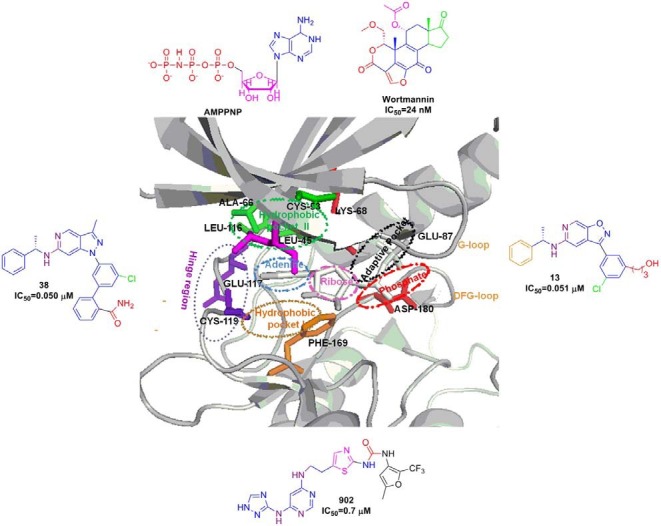
Fig. 1. Crystal structure of the ATP binding site of zPlk1 KD in complex with adenosine diphosphate (PDB ID: 3D5W) was depicted in ribbon structure with key residues. Ribbon structure depicts various binding pockets such as adenine (blue), ribose (pink), phosphate (red), hydrophobic (green and gold), adaptive pocket (black) and hinge region (violet) along with the corresponding interacting residues with stick models. Outer box shows all the reported Plk1 kinase inhibitors and the engaged atoms with the hydrogen bonding, π-π stacking etc are highlighted with corresponding color in the pockets.

