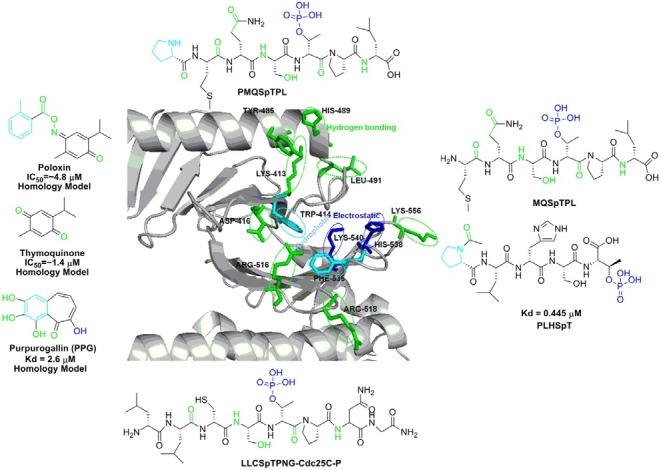
Fig. 3. Crystal structure of the phosphopeptide binding site of hPlk1 PBD in complex with PLHSpT peptide (PDB ID: 3VFH) was depic-ted with key regions indicated by different color. Outer box shows all the reported Plk1 PBD binding ligands based on their crystal structure or homology model.

