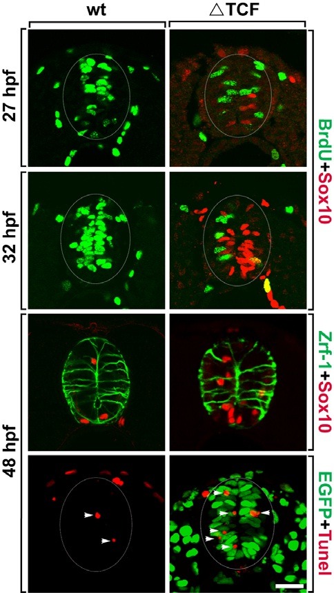
Fig. 2. Spinal precursors stop proliferation and undergo apoptotic cell death in response to ΔTcf3 expression. All images are transverse sections of the spinal cord, dorsal side up. (A-D) Double labeling of wild-type (A, C) and Tg(hsp70:tcf3-GFP) embryos (B, D) with anti-BrdU (green) and anti-Sox10 (red) antibodies. (E, F) Double labeling of wild-type (E) and Tg (hsp70:tcf3-GFP) embryos (F) with anti-Zrf-1 (green) and anti-Sox10 (red) antibodies. (G, H) TUNEL staining (red) of wild-type (G) and Tg(hsp70:tcf3-GFP) embryos (H) to detect apoptotic cell death. EGFP (green) indicates ΔTcf3 expression in response to heat-shock (H). Arrowheads indicate TUNEL+ cells (G, H). Data were obtained from 20 sections from each of five control and five heat-shocked transgenic embryos for each time point. Scale bar: 20 μm.

