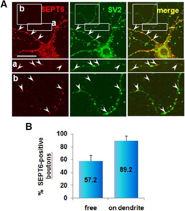
Fig. 8. Confocal microscopic images showing localization of SEPT6 at the axonal boutons. Rat hippocampal neurons in culture (DIV 21) were double-labeled with anti-SEPT6 (red) and SV2, a marker for synaptic vesicles (A) Pre-synaptic terminals on dendrites are marked with arrowheads. The free boutons en passant [box (a) and (b)] are shown at bottom in high contrast and marked with arrowhead. Statistics of boutons positive for SEPT6 is shown in percentage (B) Scale bar, 30 μm.