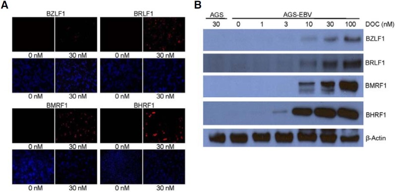
Fig. 4. Induction of EBV lytic gene by docetaxel treatment. Cells were treated with indicated concentrations of docetaxel for 72 h. (A) Expression of EBV lytic genes was analyzed by immunofluorescence assay. Anti-BZLF1 (1:50), anti-BRLF1 (1:100), anti-BMRF1 (1:200), anti- BHRF1 (1:100) antibodies, and Alexa Fluor 555-conjugated goat anti-mouse IgG (1:500, red) were used. Nuclei were stained with DAPI (blue). (B) Western blot was performed using anti-BZLF1 (1:500), anti-BRLF1 (1:500), anti-BMRF1 (1:500), anti-BHRF1 (1:250), and anti-β-actin (1:2000) antibodies.