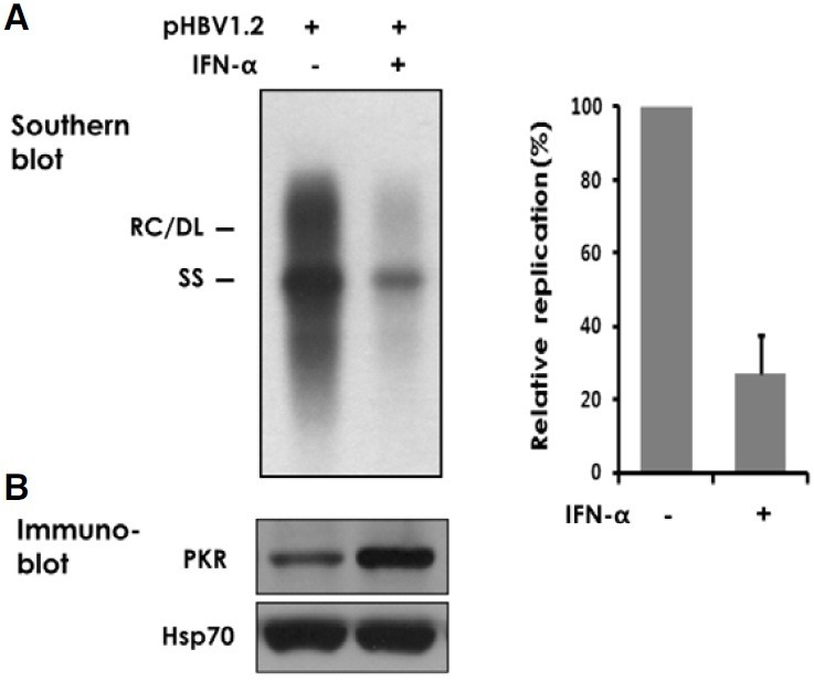
Fig. 1. Inhibition of HBV replication by IFN-α. Huh-7 cells transfected with 2 μg of pHBV1.2 were treated with 500 U/ml of IFN-α for four days. (A) Intracellular viral capsids were precipitated from cell lysate and the encapsidated viral DNA was analyzed by Southern blotting. The relaxed circular (RC), double-stranded linear (DL), and singlestranded (SS) forms of viral DNA are indicated on the left. Signal intensity of four similar experiments was quantified using TINA image analysis software and an average value was shown on the right. (B) An aliquot of cell lysate was immunoblotted for PKR. Hsp70 was used as a loading control.