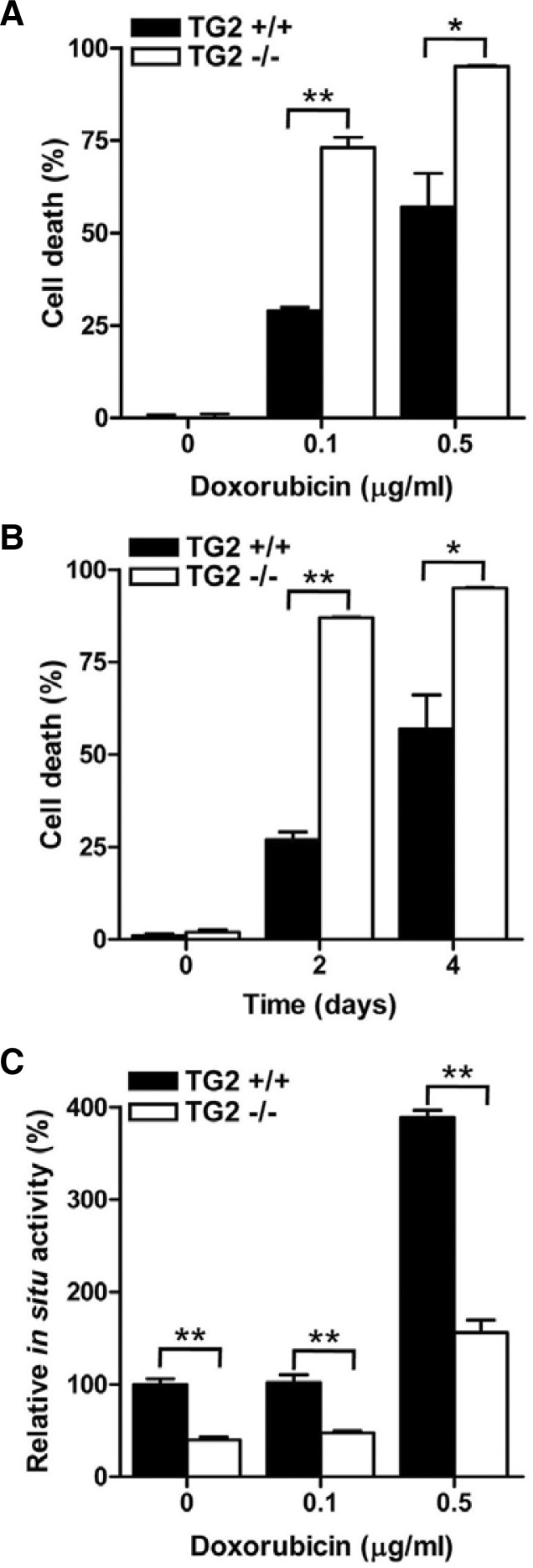
Fig. 3.
TG2 expression promotes the survival of doxorubicin-treated MEFs. (A) Wild type (TG2+/+) and TG2-null (TG2−/−) MEFs were treated with 0.1 and 0.5 μg/ml of doxorubicin for 4 days. (B) MEFs were treated with 0.5 μg/ml doxorubicin for 2 and 4 days. The extent of cell death was evaluated by trypan blue exclusion staining. Cell death was expressed as the percentage of dead cells out of the total number of cells. (C) MEFs were treated with 0.1 and 0.5 μg/ml of doxorubicin for 24 h. Each sample was analyzed for the intracellular activity of TG2. The data represent the mean values ± standard deviations based on 3 independent experiments. *, p < 0.05; **, p < 0.01.

