Fig. 6.
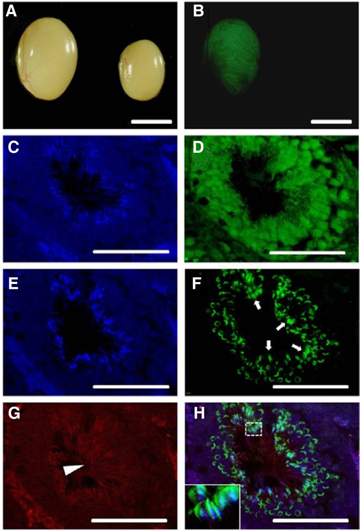
Histology of W sterile recipient testes injected with spermatogonial stem cells expres-sing lentiviral vector EF1-GFP (2 ug/ml of DOGS, MOI 5, 6 h infection) viewed under a microscope. Whole-mount images of GFP colonies from donor-derived spermatogenesis at 2 months after transplantation (A, B) and immunohistochemical evaluation of transplanted seminiferous tubules (C–H). (A, B) Light and correspond-ing fluorescence photomicrographs, left testis, each GFP expressing area indicates donor-derived spermatogenesis, right testis, control testis without transplantation did not show fluorescence; (C–H) Fluorescent image showing multiple layers of GFP-expressing donor-derived germ cells and sperm. Cross-section through a colony of donor spermatogenesis stained by GFP antibody and the incidence of sperm detected by PNA lectin and tubulin antibodies. (C–D) and (E–H) images represent a right next section from the same tubule. (C, E) DAPI, (D) Alexa flour 488 labeled GFP, (F) Alexa flour 488 labeled PNA lectin, (G) Alexa flour 647 labeled tubulin, (H) Overlay of E–G fluorescent signals. Inset displays a higher magnification of sperm heads in the dashed white box (Arrow, PNA stained acrosome of sperm; Arrow head, tubulin stained sperm tail, Scale bars = A, B: 5 mm, C–H: 50 um).

