Fig. 1.
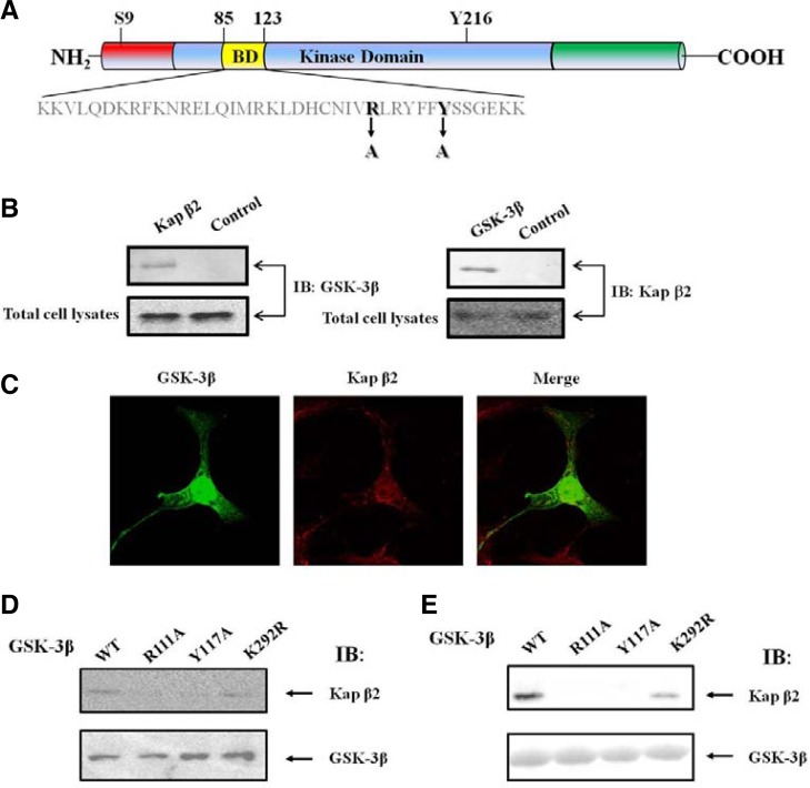
The putative PY NLS in GSK-3β and interaction between exogenous GSK-3β and Kap β2. (A) GSK-3β contains the putative-conserved Kap β2 binding motif (109IVRLRYFFY117) within its binding domain (yellow). Two point mutants were prepared in order to define the binding region. GSK-3β mutants in the Kap β2 putative binding motifs, were also prepared, and the mutated sequences were indicated as Y117A (109IVRLRYFFY 117 changes to 109IVRLRYFFA 117) or R111A (109IVRLRYFFY 117 changes to 109IVALRYFFY 117). For these mutants control, we used the unrelated GSK-3β K292R mutant. (B) Following immunoprecipitation (IP) using an anti-Kap β2 antibody, an immunoblot (IB) was performed using an antibody against GSK-3β (left). The immunoprecipitated GSK-3β complexes were applied to the immunoblot, using an anti-Kap β2 antibody (right). For the negative control, normal mouse serum was used for immunoprecipitation. (C) Confocal fluorescence micrographs showing the endogenous GSK-3β and Kap β2 in HEK293 cells. Kap β2 was visualized by immunofluorescence in fixed and permeablized cells using polyclonal antibodies to human Kap β2 or GSK-3β and Alexa Fluor 568 conjugated donkey anti-rabbit IgG or Alexa Fluor 488 conjugated mouse anti-rabbit IgG. The yellow pattern resulting from the merging of red and green colors indicates the co-localization of both proteins at a specific region of the nuclear membrane and nuclear. (D) HEK293 cells were transiently transfected with expression vectors, HA-GSK-3β WT, R111A, Y117A. Following immunoprecipitation (IP) using an anti-HA antibody, either Kap β2 (upper lane) or GSK-3β (down lane) was detected with the immunoblot (IB) using an antibody against Kap β2 or GSK-3β. (E) In vitro pull down assay with the fusion protein of GSK-3β (WT, R111A, Y117A, K292R). Whole cell lysates of HEK293 cells was incubated with 1 μg of each glutathione agarose tagged recombinant GSK-3β (WT, R111A, Y117A, K292R). The immunoblot was performed to detect Kap β2 with its antibody (upper lane). The recombinant GSK-3β (WT, R111A, Y117A, K292R) protein amount were monitored with the coomasaie blue staining (bottom lane).

