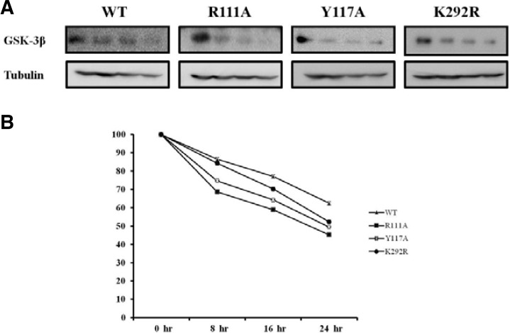
Fig. 4.
The protein stability of GSK-3β and its PY mutant. HEK293 cells (2.5 × 10⁵ cells per well) in 100 mm plates were transfected with 8.0 μg of expression vector with HA-GSK 3β wt or its PY mutant plasmid. The medium was replaced with medium containing 200 μg/ml cycloheximide 36 h after transfection (0-hr time point). Cell lysates were harvested at 0, 8, 16, and 24 h then analyzed by immunoprecipitation and Western blotting using anti-HA antibodies, and assayed in five time repeats. The relative optical density (OD) was measured by image analysis of the dried SDS-PAGE gel with the Fuji Image Quant software (Fujifilm, Japan), according to the manufacturer’s instructions.

