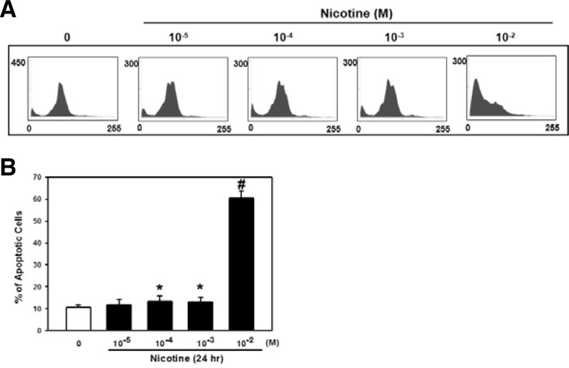
Fig. 4.
Effect of nicotine on apoptotic cell death in PDLSCs. (A) Cells were treated with various concentrations of nicotine (0–10−2 M) for 24 h and the DNA contents of cells were then measured by flow cytometry. (B) The bars denote the percentage of cells in the subG1 phase. The values reported are the mean ± S.D. of five independent experiments. *P < 0.05 or #P < 0.001 vs. control value.

