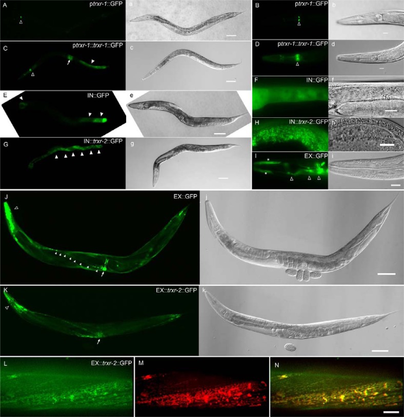
Fig. 1.
Expression pattern of trxr-1 and trxr-2 in C. elegans. A transcriptional construct ptrxr-1::gfp is mainly expressed in M2 neurons [open arrowheads in (A) and (B)]. A translational construct ptrxr-1::trxr-1::gfp is expressed in post intestine [arrowhead in (C)], vulva [arrow in (C)], and post pharyngeal bulb [open arrowheads in (C) and (D)]. Both transcriptional pIN::gfp and translational pIN::trxr-2::gfp constructs are mostly expressed in intestine [arrowheads in (E) and (G), respectively]. The transcriptional fluorescence signal is diffused (F), whereas the translational expression reveals the punctate and reticular mitochondrial morphology (H). The other tran-scriptional pEX::gfp is expressed in vulva [arrow in (J)], nerve cord [arrowheads in (J)], many head neurons [open arrowheads in (I) and (J)], and pharyngeal hypodermis cells [asterisks in (I)]. A translational pEX::trxr-2::gfp is mainly expressed in muscles including pharynx (open arrowheads) and vulva (arrow). TRXR-2::gfp signals in body wall muscle (L) were colocalized with Mitotracker staining (M), as shown in the merged image (N). A corresponding DIC image to each fluorescent image is shown in (A) to (K). Scale bar: 50 μm in (A), (C), (E), (G), (J), and (K); 10 μm in (B), (D), (F), (H), (I), and (N).

