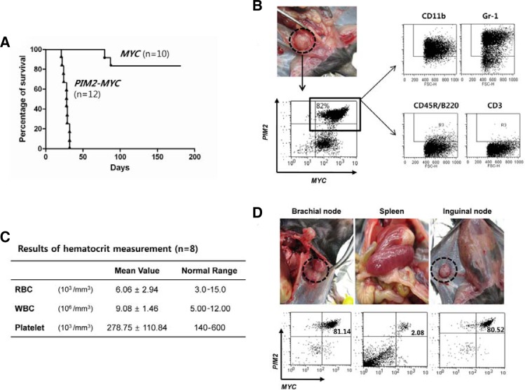
Fig. 5.
PIM2/MYC-transduced cells form myeloid sarcomas upon in vivo injection. (A) The mice received 5 × 10⁵ cells of the cell lines establised with the gene(s) indicated. Ten (MYC) and 12 mice (PIM2/MYC) were used as hosts as described in “Materials and Methods”. Eight mice from the MYC-alone group were healthy and survived for more than 150 days at which time all the mice were sacrificed. (B) A representative sarcoma that developed beneath the spine. Phenotypic analysis of sarcoma cells demonstrated the majority to still be positive for GFP and tNGFR markers and clearly of myeloid phenotype similar to the transplanted parental cells. (C) The hematocrit data for symptomatic mice are demonstrated to be within normal limits. The table shows the average hematocrit collected from eight mice that have received cells from PIM2/MYC cell line. (D) Sarcomas are formed in lymph nodes and are mostly composed of cells still expressing the markers for MYC (tNGFR) and PIM2 (GFP) expression similarly to the parental cells that were transplanted to the mice while minimal number of double-positive cells are detected in spleen. The same is true for bone marrow (data not presented).

