Fig. 5.
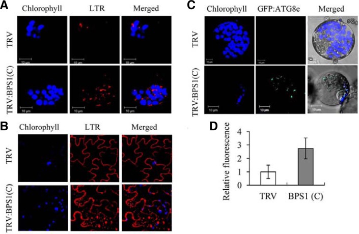
Activation of autophagy. (A) LysoTracker Red (LTR) staining of leaf protoplasts isolated from TRV and TRV:BPS1(C) lines at 15 DAI. LTR-stained punctuate autophagosome-like structures, chloro-phyll autofluorescence (pseudo-colored blue), and merged images were observed by confocal laser scanning microscopy. (B) LTR staining of leaf epidermal cells of TRV and TRV:BPS1(C) lines at 15 DAI. Note that LTR also stains the epidermal cell wall. (C) Confocal microscopy of transiently expressed GFP:ATG8e in leaf protoplasts isolated from TRV and TRV:BPS1(C) lines at 15 DAI. (D) LTR-derived red fluorescence of the protoplasts shown in (A) was quantified by confocal microscopy. Data points represent means ± SD of 30 individual protoplasts.

