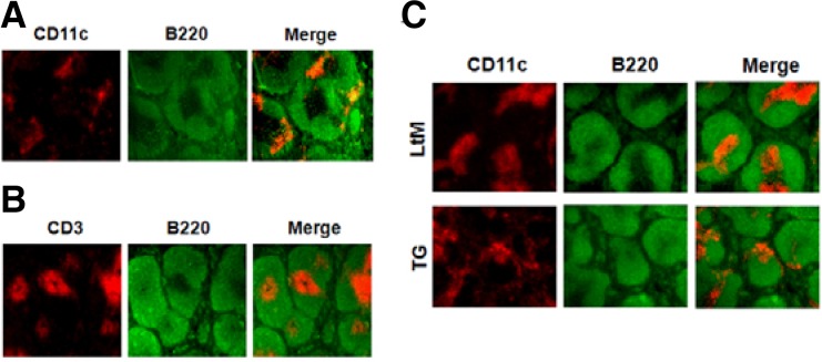
Fig. 2.
DC localization in the spleens of LtM control or p190RhoGEF TG mice after LPS injection. (A) A frozen mouse spleen section was stained with PE-conjugated anti-mouse CD11c and FITC-conjugated anti-mouse B220 Abs. When merged together, CD11c+ DCs (red) were shown scattered in the marginal zone of B cell area (green) outside of T cell zone. (B) By staining a frozen mouse spleen section using PE-conjugated anti-CD3 and FITC-conjugated anti-mouse B220 Abs, the T cell zone (red) was discriminated from the B cell area (green). (C) Frozen spleen sections were prepared from LtM control or TG mice that were injected with LPS (25 μg) for 6 h. By staining sections with PE-conjugated anti-mouse CD11c and FITC-conjugated anti-mouse B220 Abs, the localizations of DCs were compared. When merged together, the DCs (red) were stained within the T cell zone in the LtM mice. In contrast, the DCs (red) were shown scattered in the marginal zone of B cell area (green) outside of the T cell zone in the TG mice. Experiments were performed in three distinct TG lineages at least. The data shown are representative of at least six separate experiments.

