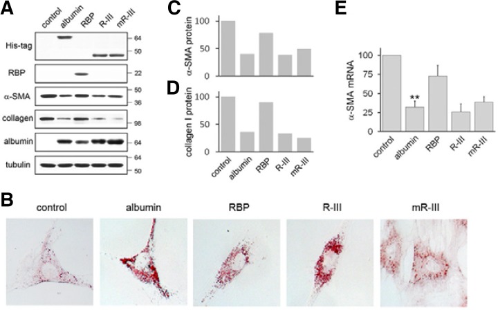
Fig. 2.
Expression of R-III deactivates HSCs. (A) HSCs after passage 2 (HSCs-P2) were transiently transfected with either empty vector (control) or expression plasmids for His-tagged albumin, RBP, His-tagged R-III or mutant R-III (mR-III), and cell lysates were analyzed by Western blotting. The Western blots are representative of three independent experiments from separate cell preparations. α-tubulin serves as loading control. (B) Transfected HSCs were subjected to oil red O staining. (C, D) Quantitative result of Western blots of α-SMA (C) and collagen type I (D) using image J software. (E) Total RNA was isolated from transfected HSCs and analyzed for α-SMA by real-time PCR. The data are expressed as the percentage of control HSCs and represent the means with standard deviation (n = 3). **P < 0.01 compared with control HSCs.

