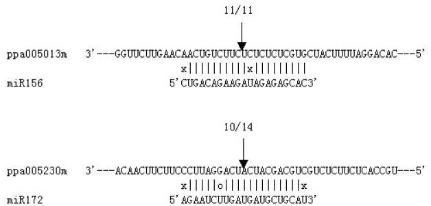
Fig. 4.

Mapping of mRNA cleavage sites by RNA ligase-mediated 5′ RACE. Each top strand depicts a miRNA-complementary site in the target mRNA, and each bottom strand depicts the miRNA. Watson-Crick pairing (vertical dashes), G:U wobble pairing (circles) and mismatched base pairing (X) are indicated. Arrows indicate the 5′ termini of mRNA fragments isolated from peach. Numbers indicate the fraction of terminating cloned PCR products.
