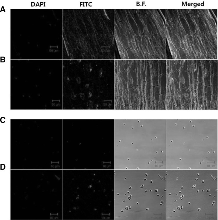
Fig. 4.
Immunolocalization of GMK1 during NaCl treatment. (A) As a control, longitudinal sections of soybean root were treated with anti-GMK1 serum, and then exposed to a FITC-conjugated antibody. (B) Soybean seedling treated with 300 mM NaCl for 60 min was longitudinally sectioned and was treated with anti-GMK1 serum and FITC-conjugated antibody. (C) Nuclei were isolated from control seedlings using PARTEC (Germany) nuclear isolation reagents and then treated with anti-GMK1 antibody and FITC-conjugated antibody. (D) Nuclei were isolated from 300 mM NaCl-treated soybean seedlings and were handled in same manner as those in (C). All images were obtained using a confocal microscope. Nucleus was seen as blue with DAPI staining. B.F. indicates bright field.

