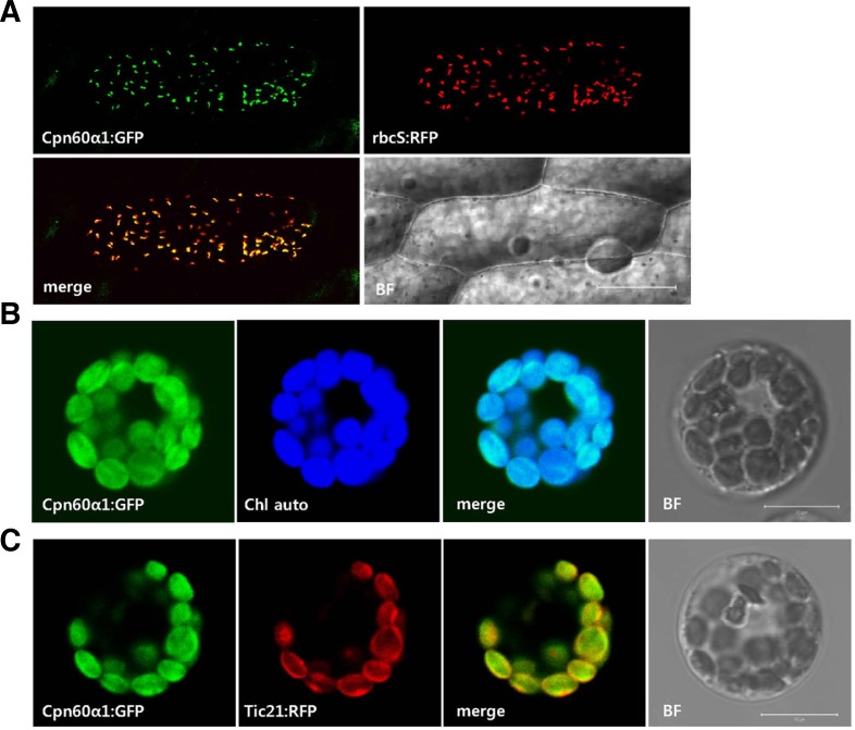
Fig. 5.
Subcellular localizations of OsCpn60α1. (A) p35S-OsCpn60α1: GFP construct was co-transfected with plasmid marker p35S-rbcS: RFP into onion epidermis cells by particle bombdardment. Scale bar = 100 μm. (B) OsCpn60α1:GFP fusion protein was expressed alone in rice mesophyll protoplasts. GFP signal was merged with chlorophyll auto-fluorescence (Chl auto). (C) p35S-OsCpn60α1:GFP construct was co-electroporated with p35SAtTic21: RFP plasmid (a chloroplast inner-membrane marker) into rice mesophyll protoplasts. BF, bright field; Scale bar = 10 μm.

