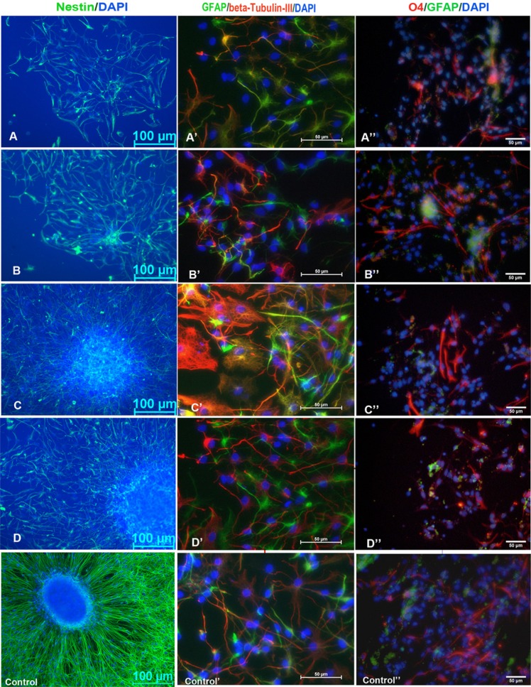
Fig. 5.
Immunofluorescence staining of cell phenotypes cultured on the amino modified surfaces and PLL surfaces. Nestin immunofluo-rescence stained for NSCs after one day of culturing; GFAP, β-tubulin III and O4 immunofluorescence double staining of NSCs differentiation showed astrocytes, neurons and oligodendrocytes after seven days of culturing. Blue indicates DAPI staining of nuclei. Scale bar (A–D), 100 μm; scale bar (A′B′C′D′ and A″B″C″D″), 50 μm. (A–D), amino modified surfaces with different densities; (Control), PLL coated surfaces.

