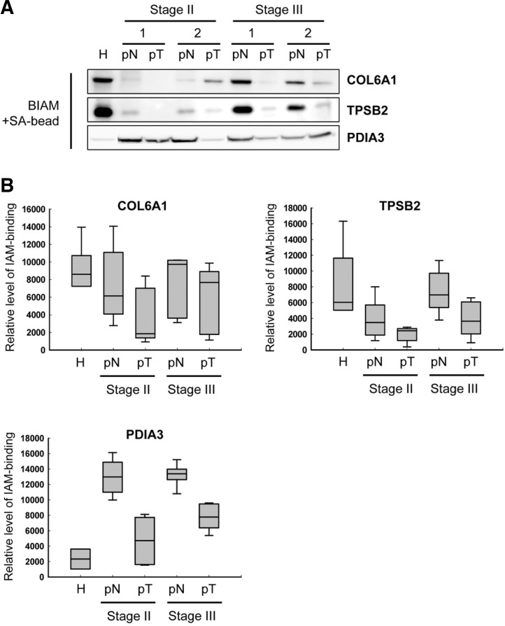
Fig. 2.
Detection of proteins containing IAM binding cysteine by immunoprecipitation using BIAM-streptavidin beads. (A) A representative gel image of IAM-binding level of COL6A1, TPSB2 and PDIA3. The cysteine oxidation of COL6A1, TPSB2 and PDIA3 was confirmed in the membrane fraction of normal subject, non-tumoral, and tumoral tissues (see the information in Table 2). Samples were run in triplicate. (B) Histograms of IAM-binidng level of COL6A1, TPSB2, and PDIA3. The graph is shown along with the numeric data obtained by densitometry analysis (n = 5). H, normal tissue of healthy subject; pN, non-tumor tissues of CRC patients; pT, tumor tissues of CRC patients.

