Fig. 4.
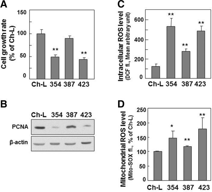
Decreased mtOGG1 expression-associated mitochondrial dysfunction may also be associated to delayed cell growth and increased intracellular ROS. Chang-L cells (Ch-L) and four different SNU hepatoma cell lines (SNU354, SNU387, SNU423) were cultured for 2 days to maintain exponentially growing state. (A) Cell growth rates were measured by counting trypan blue positive cells. No clear dead cells were observed. (B) Western blot analysis. (C) Intracellular ROS levels were monitored by flow cytometric analysis after staining cells with DCFH-DA. (D) Mitochondrial ROS levels were monitored by flow cytometric analysis after staining cells with MitoSOX fluorescence dye. **, < 0.01 vs. Chang-L cell.

