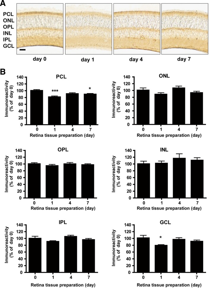
Fig. 4.
Differential Bcl-2 expression in mouse retina after single BSO administration. (A) Immunohistochemical analysis of Bcl-2 expression in mouse retina under single BSO administration condition. Immunoreactivity intensities were determined by NIH ImageJ. Scale bar represents 1 mm. (B) Immunohistochemical analysis of Bcl-2 expression in mouse retina layers under single BSO administration condition. Intensity of immunoreactivities was determined from at least 12 retina tissue sections of 8 eyeballs (4 animals). PCL, ONL, OPL, INL, IPL, and GCL represent photoreceptor cell layer, outer nuclear layer, outer plexiform layer, inner nuclear layer, inner plexiform layer, and ganglion cell layer, respectively. Statistical significances are indicated as *p < 0.05, **p < 0.01 and ***p < 0.001 (one-way ANOVA).

