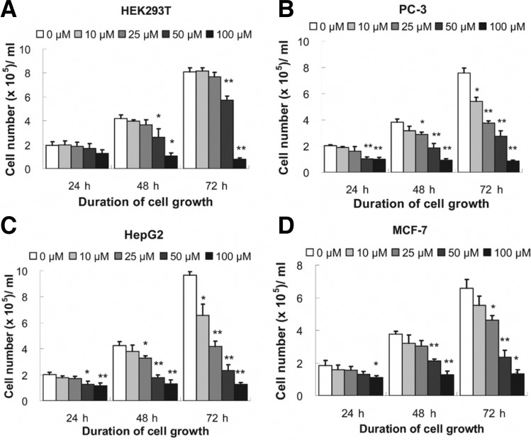
Fig. 1.
Effects of RSV on the growth of cells. HEK293T (A), PC-3 (B), HepG2 (C) and MCF-7 (D) cells. Cells (1.0 × 05/ml) were treated with 0–100 μM of RSV for 24–72 h. After harvesting, trypan blue staining was performed for cell counting. Results in bars are presented as mean ± SEM (n = 5); **P ≤ 0.01 and *P ≤ 0.05, compared with control (0 μM RSV), analyzed by one-way ANOVA and LSD post-hoc test.

