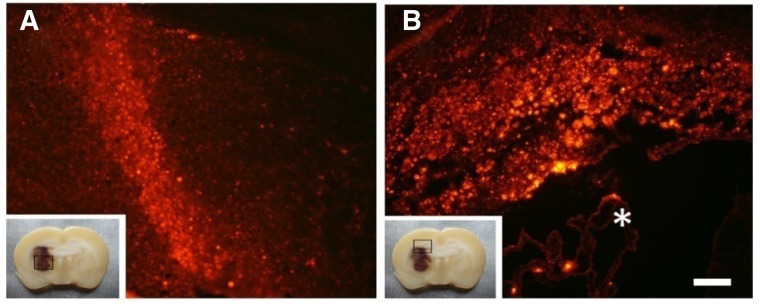
Fig. 1.
BMSCs survived and migrated in the rat brain after ICH. At 35 dpi, PKH26-labeled cells migrated into the lesion site (A) and accumulated in the corpus callosum and hippocampus (B). The box at the left lower side of each image represents low magnification of coronal slices of the ICH rat brain. Scale bar = 300 μm. *The lateral ventricle.

