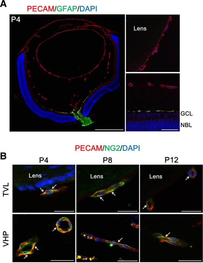
Fig. 2.
Coverage of endothelial cells in hyaloid vessels by pericytes, not by astrocytes. To figure out the presence of perivascular cells in hyaloid vessels, immunostaining of GFAP, an astrocyte marker, and NG2, a pericytes marker, was performed on the enucleated eyes from mice at P4, P8, and P12. (A) Expression of GFAP in the mouse eye at P4. Magnified views were demonstrated for comparison of the expression patterns of GFAP in between TVL (upper right) and the retina (lower right). Scale bars: 500 μm (the whole eye) and 50 μm (the magnified views). (B) Expression of NG2 in hyaloid vessels. Each image is representative of more than 50 vascular lumens of TVL and VHP, respectively. White arrows indicate distinct colocalization sites of PECAM-1 and NG2, but the immunoreactivity is not confined to the sites with white arrows. Scale bars: 50 μm.

