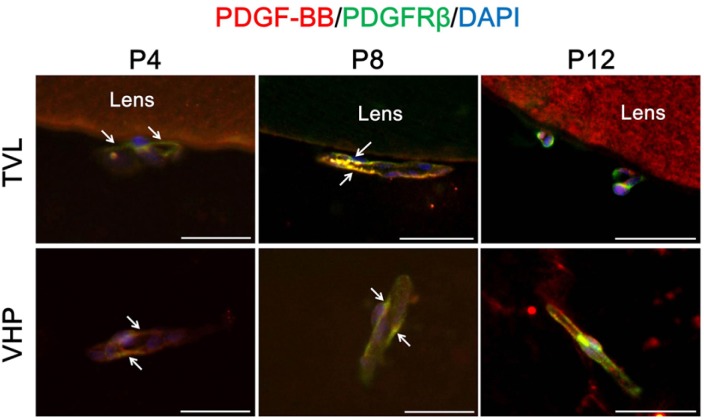
Fig. 3.
Interaction between pericytes and endothelial cells in hyaloid vessels. To figure out the interaction between two types of cells in hyaloid vessels devoid of astrocytes, immunostaining of PDGF-B and PDGFR-β was performed. Each image is representative of more than 50 vascular lumens of TVL and VHP, respectively. White arrows indicate distinct colocalization sites of PECAM-1 and NG2, but the immunoreactivity is not confined to the sites with white arrows. Scale bars: 50 μm.

