Fig. 1.
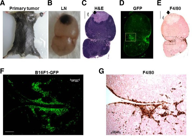
Histopathological analysis of a metastatic lymph nodes from a B16F1-GFP melanoma mouse. (A) Representative B16F1-GFP melanoma mouse with a primary tumor. (B-E) Histological analyses of a representative metastatic lymph node section: gross photograph (B), H&E staining (C), GFP fluorescence (D), and IHC staining with the F4/80 antibody (E). Scale bars, 500 μm. The arrow indicates necrotic regions of the tumor. (F-G) Magnified views of fluorescence images of the rectangular regions (F) and IHC staining of the same magnified rectangular regions (G). Scale bars, 100 μm.

