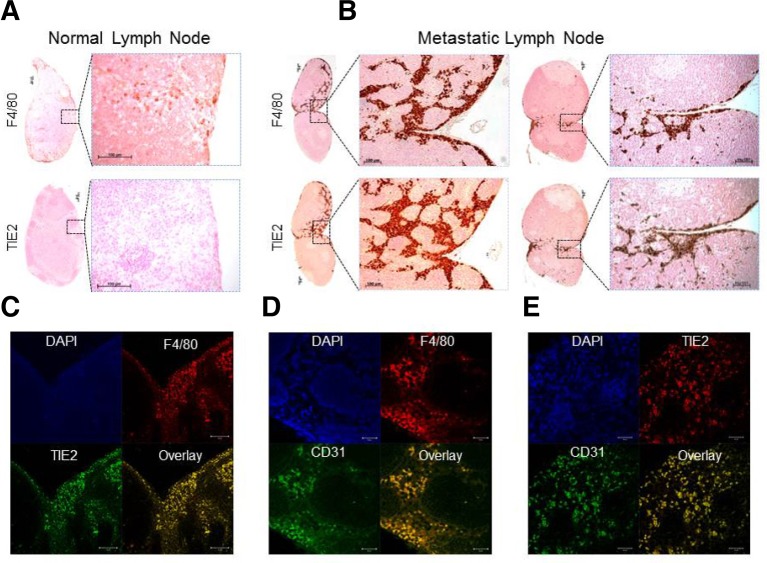
Fig. 3.
Immunological assessment of TIE2 and CD31 protein as TAM-specific markers of micro- or macro-metastasis. (A) IHC staining of serial sections from normal lymph nodes using anti-F4/80 and anti-TIE2 antibodies. Boxed regions (left) are magnified views of the right panel. Scale bars, 100 μm. (B) IHC staining of micro-meta-static lymph nodes (left) and macro-metastatic lymph nodes (right) using anti-F4/80 and anti-TIE2 antibodies. Boxed regions are shown in magnified views in the right panel. Scale bars, 100 μm. (C-E) Double immunofluorescence staining of a representative metastatic LN section using anti-TIE2 and anti-F4/80 antibodies (C), anti-F4/80 and anti-CD31 antibodies (D), and anti-TIE2 and anti-CD31 antibodies (E). Scale bars (left to right), 50, 20, and 20 μm.

