Fig. 4.
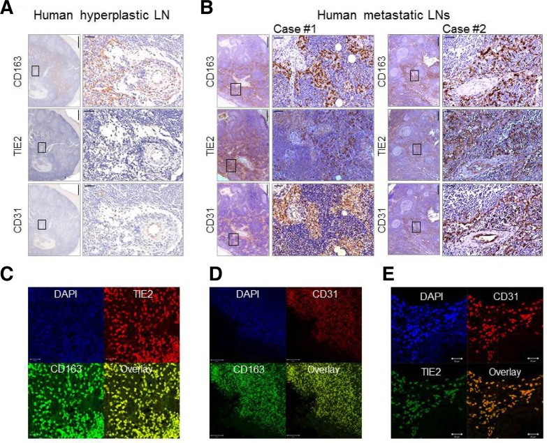
Assessment of CD163, TIE2, and CD31 expression in reactive hyperplastic lymph nodes and metastatic lymph nodes from healthy and breast tumor-bearing human subjects. (A) Immunohistochemical images for anti-CD163, anti-TIE2, and anti-CD31 antibodies are shown for representative reactive hyperplastic lymph nodes from human subjects. Boxed regions are shown as magnified views in the right panel. Scale bars, 500 μm (left) and 50 μm (right). (B) Immunohisto-chemical images for anti-CD163, anti-TIE2, and anti-CD31 antibodies are shown for metastatic lymph nodes from breast tumor-bearing human subjects. Boxed regions are shown as magnified views in the right panel. Scale bars, 500 μm (left) and 50 μm (right). (C-E) Double immunofluorescence staining of two representative metastatic lymph node sections from a breast tumor-bearing human subject using anti-TIE2 and anti-CD163 antibodies (C), anti-CD31 and anti-CD163 antibodies (D), and anti-CD31 and anti-TIE2 antibodies (E), Scale bars (left to right), 20, 50, and 20 μm.

