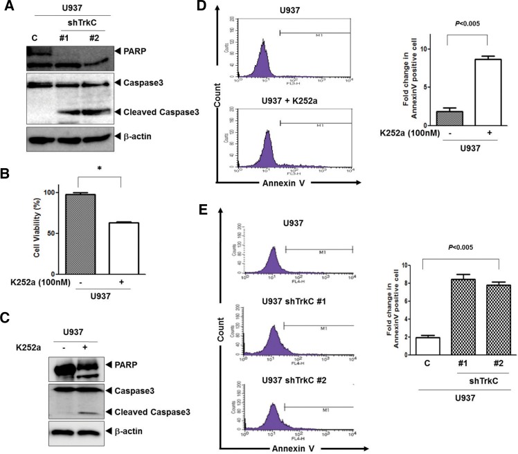
Fig. 3.
Loss of TrkC leads to loss of cell viability. (A) Cleavage of procaspase-3 and PARP of U937 control-shRNA or U937 TrkC-shRNA cells was analyzed by Western blotting. (B) Cell viability of U937 cells after treatment with K252a (100 nM) or vehicle control [dimethyl sulfoxide (DMSO)] was determined by MTT assay. Experiments were performed and the results shown reflect the mean and standard mean error (SEM) of each group. The experiments were repeated three times with similar results. *P < 0.0001 as determined by a Student’s t-test. (C) Cleavage of procaspase-3 and PARP of U937 cells with or without treatment with K252a was analyzed by Western blotting. (D) Representative FACS histograms indicating the percentage of apoptotic U937 cells after treatment with K252a as determined by binding of FITC-conjugated Annexin V. FACS analyses were conducted 24 h after culture. Right columns, mean number of Annexin V-positive cells expressed as a percentage of total cells. (E) Representative FACS histograms indicating the percentage of apoptotic U937 control-shRNA or U937 TrkC-shRNA cells as determined by binding of FITC-conjugated Annexin V. FACS analyses were conducted 24 h after culture. Right columns, mean number of Annexin V-positive cells expressed as a percentage of total cells.

