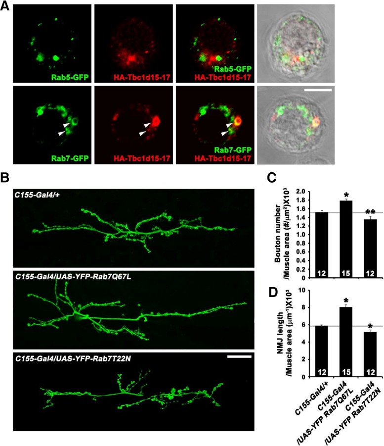
Fig. 4.
(A) Confocal images of S2 cells overexpressing both HA-Tbc1d15-17 and GFP-Rab5 or GFP-Rab7 stained with anti-GFP and anti-HA antibodies. Some vesicular structures are labeled for both Tbc1d15-17 and Rab7 (arrows). (B–D) Presynaptic Rab7 regulates synaptic growth at the NMJ. (B) Confocal images of NMJ 6/7 labeled with anti-HRP in C155-GAL4/+, C155-GAL4/UAS-YFP-Rab7Q67L, and C155-GAL4/UAS-YFP-Rab7T22N third instar larvae. (C, D) Quantification of bouton number (C) and NMJ length (D) at NMJ 6/7. *P < 0.001. Scale bars, 20 μm.

