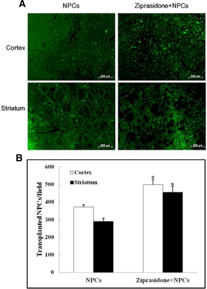
Fig. 3.
Survival rate of transplanted NPCs. (A) Immunohistochemistry showed GFP positive cells (green) in the cortical and striatal ischemic boundary zone at 7 days after MCAO (50×). (B) Quantitative analysis of the number of transplanted NPCs. GFP positive expression was significantly increased in the combination of ziprasidone and NPCs treated group compared with the individual NPCs group. Data are the mean ± S.E.M. (n = 3) *p < 0.05 vs. NPCs group.

