
Fig. 6.
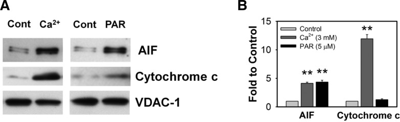
PAR polymer induces AIF release from isolated mouse brain mitochondria. (A) Representative immunoblot images of AIF and cytochrome c release. Isolated mouse brain mitochondria were incubated with Ca2+ (3 mM) o r PAR polymer (5 μM) for 30 min at 37°C in the KCl-based assay buffer containing ATP (3 mM), ADP (0.8 mM), succinate (5 mM) and rotenone (2 μM). Released levels of AIF or cytochrome c in the supernatant of the reaction mixture were analyzed by immunoblotting. Mitochondrial pellets were probed with anti-VDAC-1 antibody to confirm that equal amounts of mitochondria were used in the experiments. (B) Quantitative analysis of immunoblot images by densitometric analysis. Immunoblot results were analyzed by ImageJ software. Bar represents fold-increase of released AIF or cytochrome c to control (n = 3, mean ± SEM). Significant difference to control was determined by paired t-test (**P < 0.01).