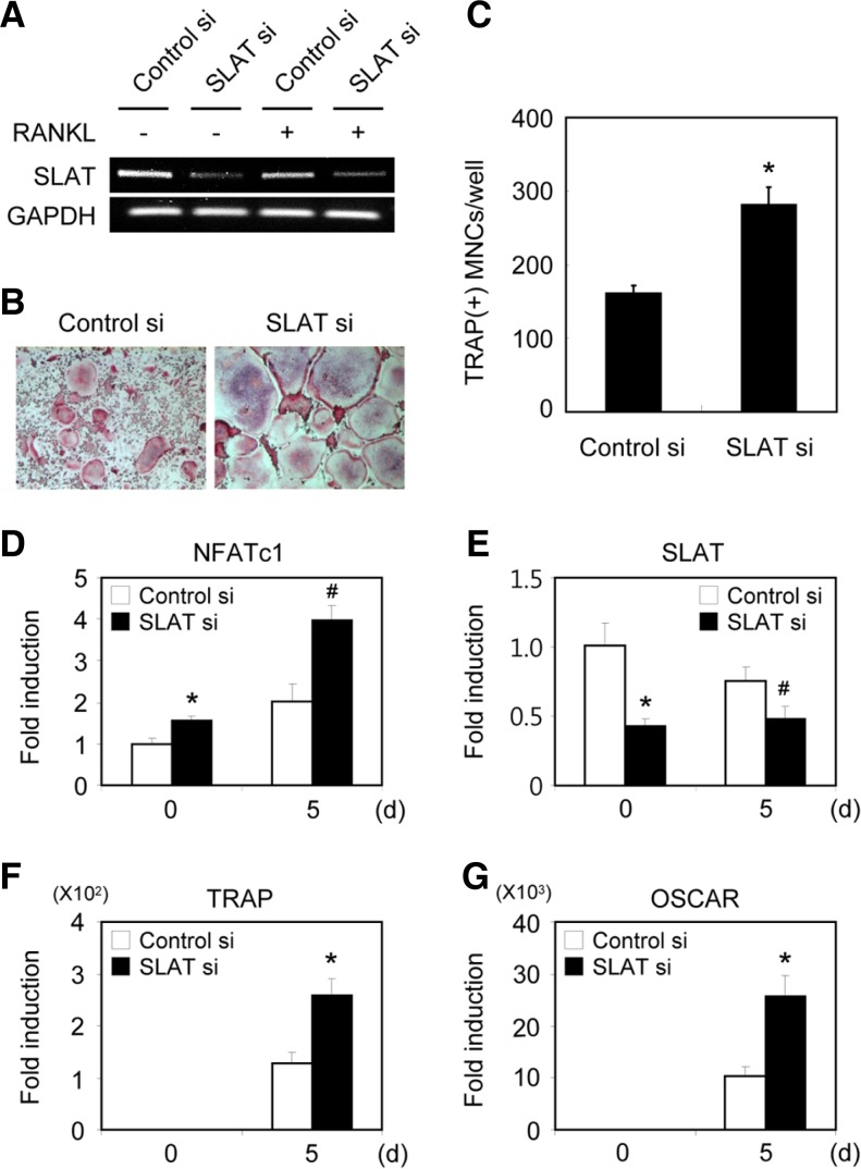
Fig. 3.
Silencing of SLAT enhances osteoclast formation and induction of NFATc1. (A) BMMs transfected with GFP or SLAT siRNA were cultured for 2 days in the absence or presence of RANKL. RTPCR was performed. (B) BMMs transfected with GFP or SLAT siRNA were cultured for 5 days in the presence of M-CSF and RANKL. Cultured cells were fixed and stained for TRAP. (C) TRAP(+) MNCs were counted as osteoclasts. (D-G) BMMs transfected with GFP or SLAT siRNA were cultured with M-CSF and RANKL for the indicated times. Real-time PCR analysis was performed with the primers specific for SLAT, NFATc1, OSCAR, TRAP, and GAPDH (control). #P < 0.05, *P < 0.01 vs control.

