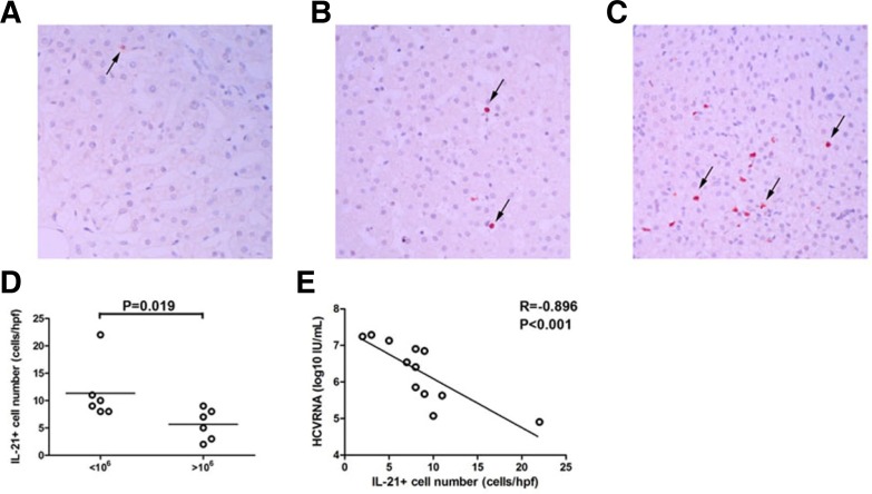
Fig. 2.
IL-21+ cells accumulated in the livers of CHC patients with a low viral load. Immunohistochemical staining for IL-21+ cells in situ in the liver of (A) healthy control (n = 1) (magnification, 200×) and CHC patients with (B) a high viral load (n = 6) and (C) a low viral load (n = 6) (magnification, 200×). Positive cells stained red, as indicated by the arrows. (D) Numbers of IL-21+ cells in CHC patients with a low viral load and a high viral load. Horizontal bars represent the median numbers of IL-21+ cells. One dot represents one individual. (E) Correlations between IL-21+ cells and plasma HCV RNA in CHC patients. One dot represents one individual. P-values are shown.

