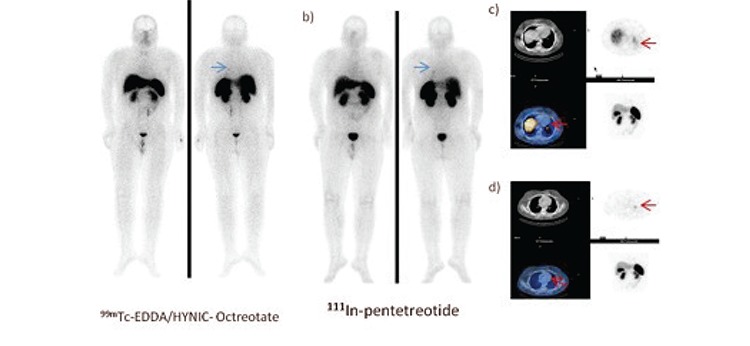
Figure 3. 18-year-old man with the diagnosis of ectopic Cushing’s syndrome a) 99mTc- EDDA/HYNIC-Octreotate, b) 111In-pentetreotide images of SRS. Metastaticlymph node seen at the left hilar area but primary focus was not detected in the planar images. c) SPECT and SPECT-CT fusion images of the 99mTc-EDDA/HYNIC-Octreotate revealed a foci of pathological tracer uptake in the lower lobe of the left lung (arrows) d) The left hilar lymph node involvement wasseen on SPECT and SPECT-CT fusion images of the 99mTc- EDDA/HYNIC-Octreotate.

