
Figure 4.
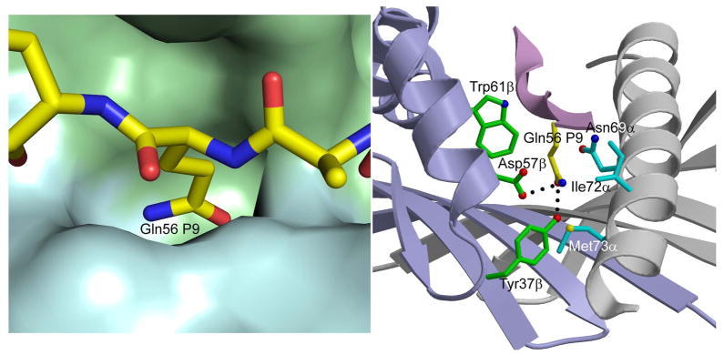
Interaction of P9 Gln56 with HLA-DR4. Left panel, molecular surface of HLA-DR4 (MHC α-chain, cyan; MHC β-chain, green) showing the P9 pocket that accommodates the side chain of P6 Gln56. Right panel, contact residues of HLA-DR4 are drawn and labeled. Hydrogen bonds are indicated by broken black lines.