
Figure 5.
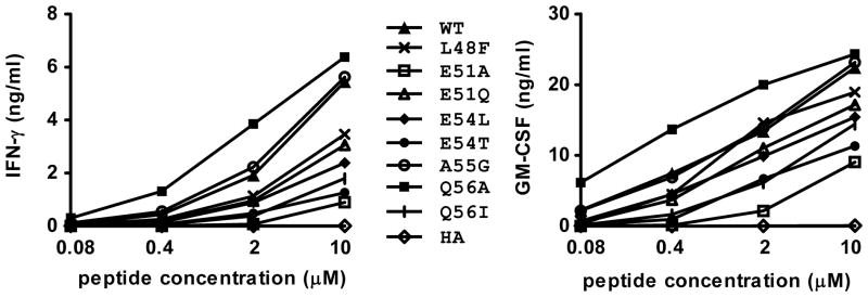
Q56A substitution at the P9 anchor residue of gp10044–59 confers enhanced recognition by the G7 clone. HLA-DR4+ 1102-EBV cells were pulsed with gp100 peptides at the indicated concentrations for 24 hrs. Then G7 cells were added and cultured overnight. Supernatants were harvested and tested for IFN-γ (left) and GM-CSF (right) secretion by ELISA. T cell recognition of the Q56A was enhanced approximately 5-fold compared to WT gp100 peptide. APLs not shown in this figure did not stimulate detectable cytokine secretion, including Y49M (P2 substitution), P50A (P3), W52A (P5) and T53V (P6). Results representative of 3 experiments. HA, influenza hemagglutinin307–319, an HLA-DR4-restricted peptide that was used as a negative control. WT, wild type gp10044–59.