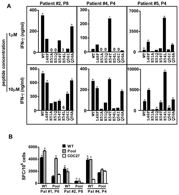
Figure 7.
T cells stimulated in vitro with gp10044–59 WT peptide show selective recognition of a variety of APLs. (A) T cells were derived from melanoma patients receiving a gp100 WT peptide vaccine. Results of IFN-γ ELISAs are shown. Background values for IFN-γ secretion by T cells stimulated with APC + HA307–319 have been subtracted from data shown, as follows: Patient #2, 213 pg/ml (1 μM peptide stimulation) and 208 pg/ml (10 μM peptide stimulation); Patient #4, 187 and 191 pg/ml; Patient #5, 346 and 388 pg/ml. (B) A peptide pool including APLs was not superior to the WT peptide in raising or detecting gp100-specific immunity. Reactivity against gp10044–59 in 10-day IVS cultures was determined by IFN-γ ELISPOT assays. CD4+ T cells were stimulated with 20 μM WT gp100 or peptide pool (WT, L48F, E54L E54T and Q56A) in both IVS cultures and ELISPOT assays. CDC27, an unrelated HLA-DR4-restricted peptide, was used as a negative control. P4, P6, P8, PBMCs collected from patients post 4, 6 or 8 vaccinations. SFC, spot forming colonies, mean± SEM. WT, wild type gp10044–59. * P<0.02, × P<0.05, one-tailed t test compared to HA or CDC27 control peptides.

