Figure 4.

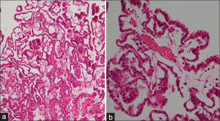
Low power (a) and high power (b) photomicrograph reveal tumor cells arranged in multiple papillae with fine fibrovascular core

Low power (a) and high power (b) photomicrograph reveal tumor cells arranged in multiple papillae with fine fibrovascular core