Figure 3.

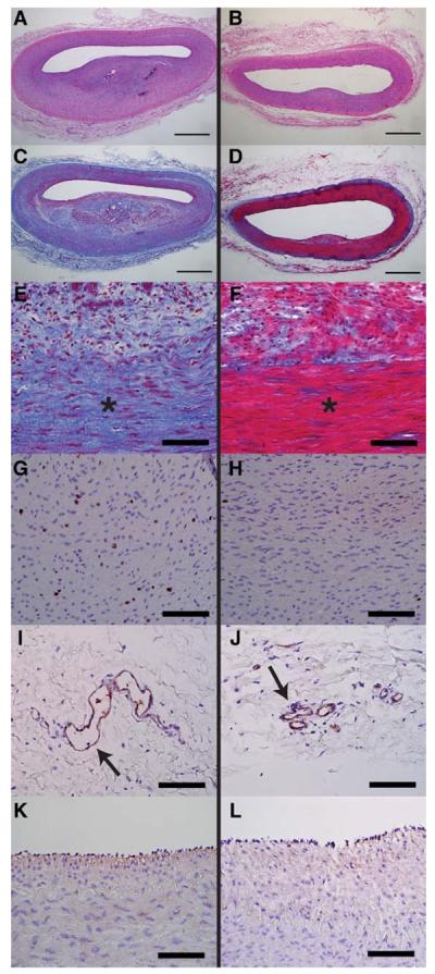
Representative histology images of pig femoral arteries 28 days after balloon angioplasty injury and adventitial injection in control (left) or nab-rapamycin (right) groups. Control arteries (A) had increased neointimal hyperplasia and luminal stenosis on hematoxylin and eosin staining compared with nab-rapamycin–treated arteries (B). Low- and high-power views of Masson trichrome stained control (C and E) and nab-rapamycin–treated arteries (D and F) demonstrate increased medial fibrosis (blue staining) in control arteries. Control arteries demonstrated more medial cell proliferation (Ki-67–stained cells in brown; G) than nab-rapamycin–treated arteries (H). Control arteries (I) had significantly more adventitial microvessels (arrow, Factor VIII staining) than nab-rapamycin–treated arteries (J). There was no significant difference endothelialization between control (K) and nab-rapamycin–treated (L) arteries (Factor VIII staining). *Marks media. Bar, 1 mm in A–D. Bar, 100 μm in E–L.
