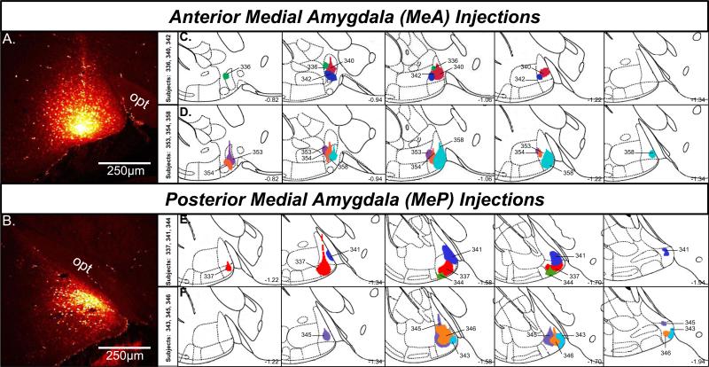
Figure 1.
A,B, Confocal image showing Fluoro-Ruby injection sites in the anterior (A) and posterior (B) medial amygdala. C,D, Schematic reconstructions of coronal sections showing the extent of individual Fluoro-Ruby injections in female mice targeting the anterior (n=6) medial amygdala. Top row: subjects 336, 340, 342; Bottom row: subjects 353, 354, 358. E,F, Schematic reconstructions of coronal sections showing the extent of individual Fluoro-Ruby injections in female mice targeting the posterior (n=6) medial amygdala. Top row: subjects 337, 341, 344; Bottom row: subjects 343, 345, 346. Sections are ordered sequentially from anterior (left) to posterior (right), with the numbers shown on the bottom right of each plate representing the distance in mm posterior to bregma. Colored regions within each plate represent individual injections, identified by the animal code. Adapted from Franklin and Paxinos (Franklin & Paxinos, 2008). MeA, anterior medial amygdala; MeP, posterior medial amygdala; opt, optic tract.

