
Figure 1.
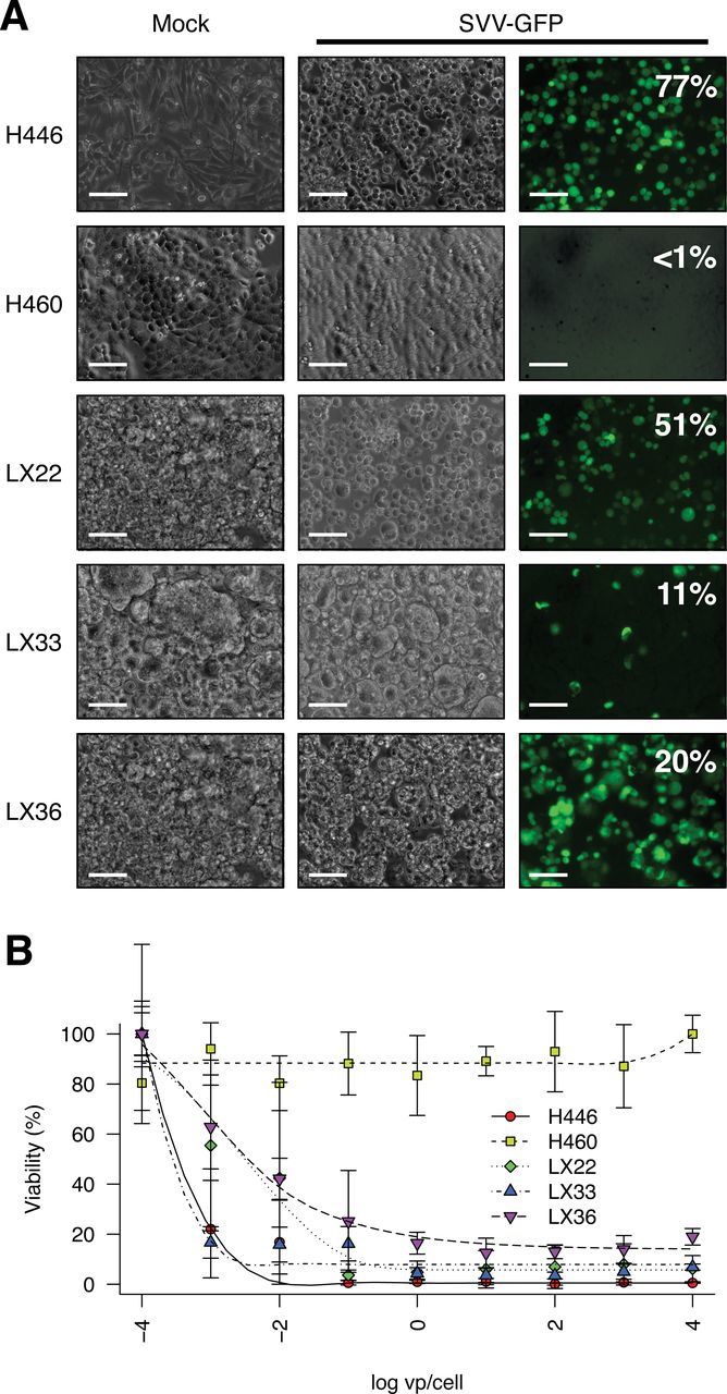
In vitro analysis of Seneca Valley virus (SVV-001) permissivity in cell lines derived from SCLC heterotransplant models. SVV-001 was serially diluted and administered from frozen stocks provided by Neotropix, Inc (Malvern, PA) as previously described (10). SVV-GFP is a derivative of SVV-001 expressing green fluorescent protein (18). Cell lines were plated in Opti-MEM containing 1 vp/cell SVV-GFP in 96-well plates. After 6 to 8 hours, cells were imaged by epifluorescence microscopy or manually triturated and analyzed by flow cytometry on a FACSCalibur (BD Biosciences). A) Fluorescence microscopy of H446 (positive control), H460 (negative control), and cell lines derived from LX22, LX33, and LX36 infected with SVV-GFP reporter virus demonstrates a high fraction of infected cells. Scale bars = 100 µm. B) The half-maximal effective concentration of wild-type SVV-001 was determined for each cell line by conversion of 3-(4,5-dimethylthiazol-2-yl)-5- (3-carboxymethoxyphenyl)-2-(4-sulfo-phenyl)-2H-tetrazolium) as previously described (10). Points represent mean values of five independent wells and uncertainty is summarized by standard deviation. Data shown are representative of four independent experiments.