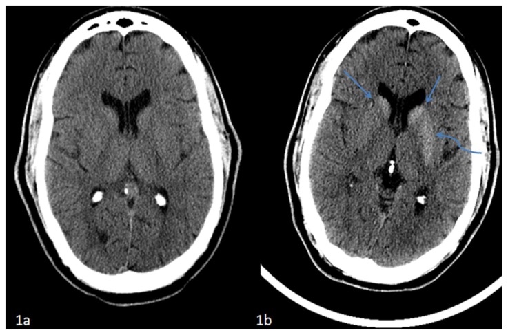
Figure 1.
42 year-old male who presented with vague right sided stroke-like symptoms and was found to have nonketotic hyperglycemia. FINDINGS: (1a) Axial non-contrast computed tomography of the head performed at ten months prior to current presentation demonstrates no abnormalities at the level of the basal ganglia. TECHNIQUE: Philips 64-slice CT scanner, 501mAs, 120 kvp, 5mm slice thickness. FINDINGS: (1b) Axial non-contrast computed tomography of the head performed at time of current presentation demonstrates interval development of hyperdensity within the caudate nuclei (straight arrows) and left lentiform nucleus (curved arrow) which respects the neuroanatomic boundaries of the basal ganglia without associated edema. TECHNIQUE: Philips 64-slice CT scanner, 350mAs, 120 kvp, 5mm slice thickness.

