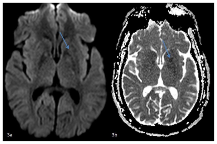
Figure 3.
42 year-old male who presented with vague right sided stroke-like symptoms and was found to have nonketotic hyperglycemia. FINDINGS: (3a) Axial DWI MR image of the brain at the level of the basal ganglia and (3b) axial ADC map correlate demonstrate faint restricted diffusion confined to the left putamen. TECHNIQUE: (3a) 1.5T Philips Intera MR scanner, diffusion-weighted imaging (DWI), TR=3873.32, TE=73.10, no contrast and (3b)1.5T Philips Intera MR scanner, diffusion-weighted ADC map, TR=3873.32, TE=73.10, no contrast.

