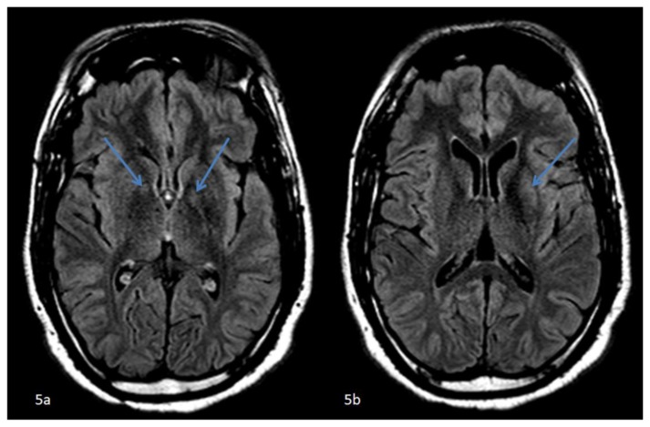
Figure 5.
42 year-old male who presented with vague right sided stroke-like symptoms and was found to have nonketotic hyperglycemia. . FINDINGS: Axial FLAIR MR images of the brain at the level of the basal ganglia (5a) slightly caudal to (5b) demonstrate subtle hyperintense signal abnormalities in the globus pallidi (left greater than right) (arrows). (5b) FLAIR hyperintense signal abnormality confined to the left putamen (arrow). TECHNIQUE: 1.5T Philips Intera MR scanner, fluid attenuated inversion recovery imaging (FLAIR), TR=11000, TE=140, no contrast.

