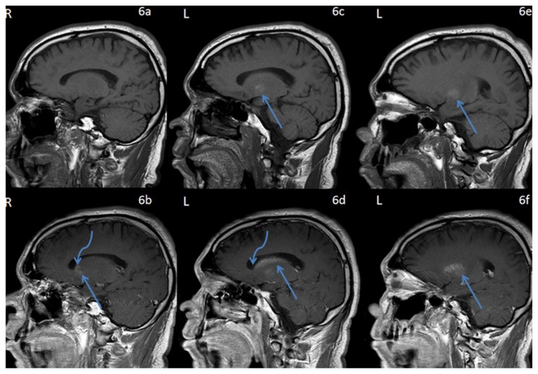
Figure 6.
42 year-old male who presented with vague right sided stroke-like symptoms and was found to have nonketotic hyperglycemia. FINDINGS: Sagittal T1-weighted pre-contrast MR images of the brain through the basal ganglia and caudate nuclei (6a) right and (6c,6e) left demonstrates intrinsic T1 shortening confined to the left globus pallidus (arrows). TECHNIQUE: 1.5T Philips Intera MR scanner, T1-weighted imaging, TR=541.53, TE=12, pre-contrast. Sagittal T1-weighted post-contrast MR images of the brain through the basal ganglia and caudate nuclei, (6b) corresponding to (6a) on the right and (6d,f) corresponding to (6c,e) respectively on the left demonstrate contrast enhancement which respects the neuroanatomic boundaries of the lentiform (straight arrows) and caudate nuclei (curved arrows) (left much greater than right). TECHNIQUE: 1.5T Philips Intera MR scanner, T1-weighted imaging, TR=541.53, TE=12, with 18ml of gadodiamide (Omniscan, GE health care, Princeton, NJ) injection.

