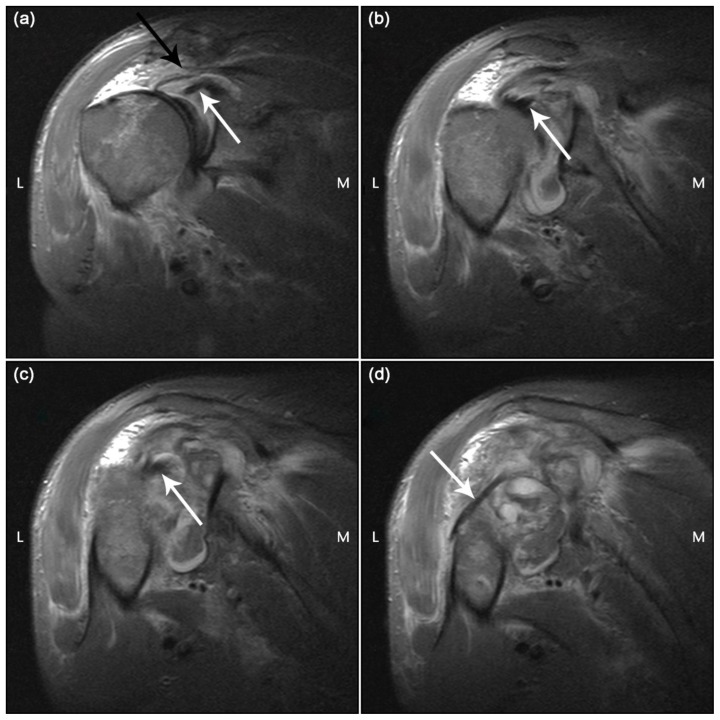
Figure 4.
69 year old male with traumatic anterior glenohumeral dislocation with failed reduction after multiple attempts, complete rotator cuff tear, posterior dislocation long head of biceps tendon and haemarthrosis.
Findings:
a) The long head of biceps tendon can be seen at its origin on the superior glenoid (white arrow).There is a full-thickness tear of supraspinatus with retraction (black arrow).
b) The intra-articular portion of the long head of biceps tendon is seen (white arrow).
c) The long head of biceps tendon is lying posteriorly with respect to the bicipital groove (white arrow).
d) The long head of biceps tendon can be seen dislocated posterior to the humerus (white arrow).
Technique: Coronal oblique STIR anterior to posterior a–d
1.5T; TR/TE/time to inversion: 4290ms/29ms/130ms; FOV 160; matrix 394 × 512; slice thickness 4 mm

