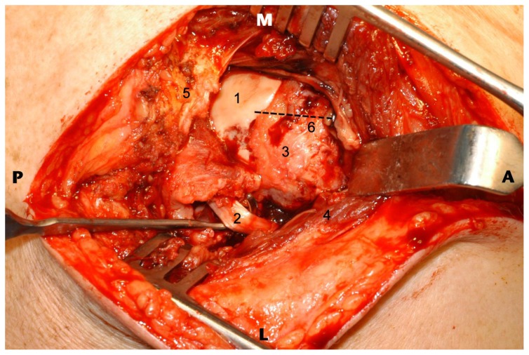
Figure 6.
69 year old male following traumatic anterior glenohumeral dislocation with complete rotator cuff tear, posterior dislocation long head of biceps tendon and haemarthrosis at MRI. Intra-operative image seen as if from above, pre-reduction following removal of the haemarthrosis where tenotomy of long head of biceps tendon and rotator cuff repair were subsequently performed.
Findings:
1) The exposed articular surface of the humeral head secondary to rotator cuff tear is seen. The humeral head remains anteriorly translated relative to the glenoid (and long head of biceps origin).
2) Posteriorly displaced long head of biceps tendon is shown by the Langenbeck retractor.
3) Supraspinatus footprint region.
4) Deltoid muscle (reflected during exposure)
5) Acromion is here.
6) Line of the bicipital groove and hence anatomical position of long head of biceps tendon.
Technique: Superior strap approach with deltoid split.

